Iliotibial band syndrome or “runner’s knee”.
Iliotibial band syndrome or “runner’s knee”.
 The iliotibial band is a connective tissue structure formed by the aponeurosis and tendon of the gluteus maximus and tensor fascia lata. This band originates in the hip (iliac blade) and runs along the lateral part of the thigh until it reaches the lower part of the knee (anterolateral side of the tibia).
One of the most frequent pathologies in this area is the iliotibial band syndrome, an overuse injury due to the repetitive rubbing of the distal area of this structure with the outer part of the knee, specifically with the external femoral condyle.
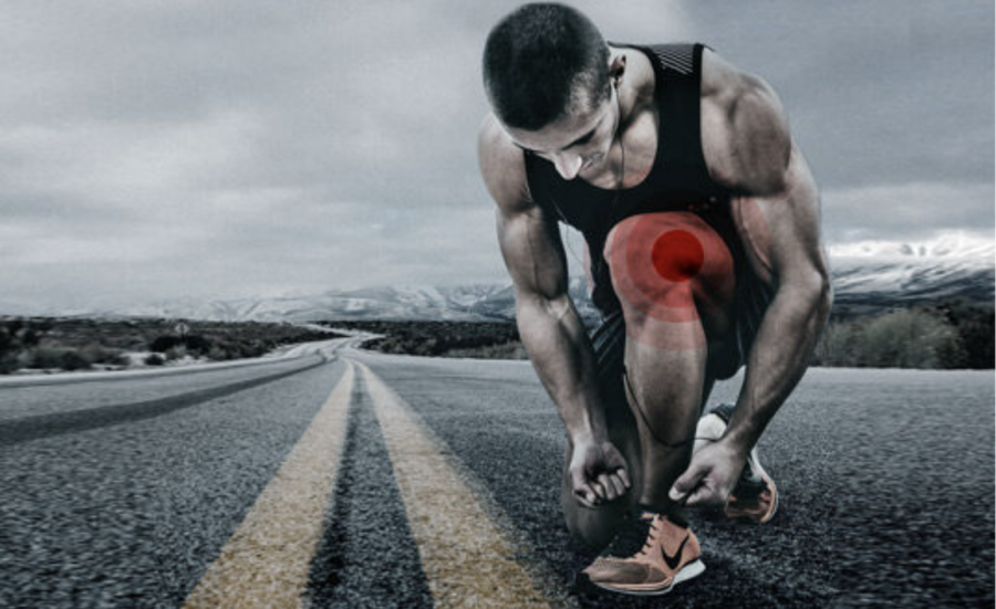
It is a characteristic injury in long distance runners and to a lesser extent in cyclists. The symptoms usually reported by these athletes are mainly pain and inflammation in the affected area, which appears at a specific running distance forcing the cessation of the activity. It generally develops progressively, with the pain appearing at shorter and shorter distances. Symptoms often appear after 15-20 minutes of running, and are aggravated on slopes and at slower paces due to a longer contact time of the iliotibial band with the external femoral epicondyle. The pain generally appears during the initial contact phase, a phase in which an eccentric deceleration contraction occurs, generating tension towards the iliotibial band.
Causes
There is no consensus as to the exact origin of the pathology, as the etiology of iliotibial band syndrome is mainly multifactorial.
Due to the frequency of this injury in runners, it suggests that continuous running is a predisposing risk factor for this pathology, since the injury mechanism is reproduced repeatedly during running.
 There are also a number of biomechanical factors that make the runner susceptible to developing this syndrome due to the increased friction of the webbing with the external femoral condyle, such as:
- Muscle shortening of hip flexors and tensor fascia latae muscle.
- Muscle imbalances. Weakness in the hip abductor muscles (specifically the gluteus medius) will lead to over-tightening of the fascia lata.
- Genu varo.
- Excessive pronation or poor foot biomechanics (tendency to flatfoot).
- The difference in leg length (dysmetria).
- Very prominent external femoral epicondyle
- Capsulo-ligamentous hyperlaxity.
Other predisposing factors are the use can be over-training, failures in the programming of the same and the use of inadequate footwear.
Symptoms of iliotibial band syndrome
The main symptoms of iliotibial band syndrome consist of pain on the outside of the knee, more specifically in the lateral epicondyle of the femur (where the band rubs), which may also be accompanied by swelling in the area. This pain appears at a specific running time/distance and gradually worsens causing even the cessation of activity. After a period of rest the pain usually subsides, appearing again when resuming running.
The pain is usually aggravated when running downhill where the work of the fascia lata is greater. In some very aggressive cases, there may even be a protrusion of the rib above the epicondyle, emitting a snapping sound.
Diagnosis
 The patient’s clinical history and symptoms will be fundamental to suspect a possible iliotibial band syndrome.
In the examination on the examination table we will confirm the diagnosis by localizing the pain by palpation of the lateral epicondyle of the femur on the external aspect of the knee. There are usually trigger points along the fascia lata and possible shortening, so we perform the Ober test.
The diagnosis can be completed with various assessment tests using surface electromyography (EMG) to see if there is correct muscle activation. Imaging tests can provide us with a lot of information on the tissue that is producing the symptomatology, both ultrasound and magnetic resonance imaging can show whether there is really tissue damage.
Treatment
The main therapeutic objective will be to reduce pain and inflammation, and then correct possible compensations, achieving a correct movement pattern and optimal muscle activation. It will be essential to continue with muscle strengthening exercises to avoid relapses.
In the conventional treatment approach we find techniques such as manual therapy, therapeutic exercise, stretching, magnetotherapy, cryotherapy, electroanalgesia, laser… among others.
An essential part will be the modification of the sport activity, adapting temporarily those activities that generate pain until it is resolved or diminished. Also take into account a progressive return to physical activity, making a correct planning of the training load, identifying and correcting those errors that caused the injury.
Therefore, it is very important to carry out Functional Rehabilitation with a professional, so that a muscle assessment by electromyography is carried out to identify and correct those movement patterns and erroneous muscle activation, as well as to develop a personalized strengthening program in which the strengthening of muscles such as the gluteus medius is a priority.
As for Advanced Physiotherapy, we can use techniques such as EPI for the regeneration of the affected muscle fibers, microEPI to reduce inflammation and Neuromodulation to reduce pain in the affected area and/or facilitate muscle activation. We can also use INDIBA to promote activation at the cellular level and normalize the tissue.
Treatment is effective if started early and should be continued until total disappearance of the discomfort.
In some cases, medical therapy such as corticosteroid infiltration or other invasive techniques may be considered. If all of the above fails, in cases where there is a very fibrotic webbing, surgical intervention is indicated.