
Chronic Chest Pain: When the Cause Lies Where No One Looks
Published on:
Last modified:
The costovertebral joints are a source of deep chest pain that is often overlooked in conventional diagnostic workups. Find out what they are, why they are overlooked, and what options are available today beyond surgery.
What are the costovertebral joints?
The thoracic spine—the region of the back that articulates with the ribs—is not just a stack of vertebrae. Each rib attaches to the spine via two distinct synovial joints, and inflammation or degeneration of these joints can cause deep, persistent, mechanical chest pain that worsens with movement, deep breathing, or changes in posture.
Costvertebral joints proper
They connect the head of each rib to the adjacent vertebral bodies. These are small synovial joints that are highly susceptible to joint degeneration (osteoarthritis).
Costotransverse joints
They connect the costal tubercle to the transverse processes of the thoracic vertebrae. Together with the former, they form the costvertebral functional unit.
Thoracic facet joints
Located between the posterior vertebral arches, they are another common source of mechanical thoracic pain that is treated in conjunction with the aforementioned causes.
Why Are They Overlooked?
Their small size and the fact that they are difficult to visualize on standard X-rays mean they are often overlooked in routine diagnostic workups. They require high-resolution MRI and targeted ultrasound examination.
Jiří Mastalír: From Prague to Barcelona in Search of a Solution
A case like Jiří’s perfectly illustrates the situation faced by many patients with chronic chest pain: years of tests and treatments that provided only partial relief and no lasting solution because the true cause of the problem had not been identified.
Years of mechanical chest pain in the Czech Republic
Despite numerous tests and treatments in his home country, Jiří had not achieved lasting relief. The actual cause remained unknown.
MRI diagnosis: bilateral costovertebral osteoarthritis at T4–T7
His most recent chest MRI revealed moderate bilateral osteoarthritis of the facet and costovertebral joints between T4 and T7, a source of deep pain that is often underdiagnosed.
Radiofrequency ablation proposal rejected due to the patient’s age
Given his young age, Jiří wanted to explore orthobiological options first before undergoing an ablative procedure. Months of independent research led him to discover our scientific publication.
Treatment at MIVI Salud – Sagrat Cor University Hospital, Barcelona
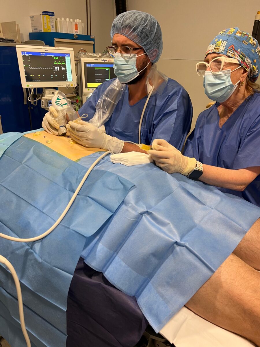
She traveled from Prague to undergo a comprehensive functional treatment of the thoracic spine—including the facet, costotransverse, and costovertebral joints from T4 to T7—using leukocyte-rich PRP under real-time ultrasound guidance, performed by our Unit Coordinator, Guilherme Ferreira, along with Dr. Gisela Roca.
Telemedicine follow-up from your home country
Jiří has already returned to the Czech Republic and is continuing his recovery with regular follow-ups through our telemedicine system.
Jiří’s case is not unique: there are many patients with chronic chest pain whose actual cause has not been identified because the costovertebral joints are rarely included in the standard diagnostic and therapeutic protocol. The combination of high-resolution imaging, interventional ultrasound expertise, and orthobiology now opens up a significant therapeutic avenue for this patient population.