Patellofemoral syndrome: What is it, what are its degrees and how can we deal with it?
Patellofemoral syndrome
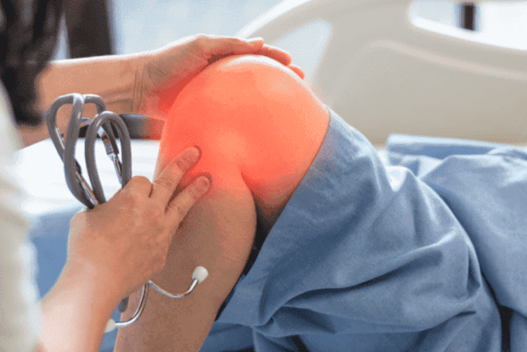
Patellofemoral syndrome, also known as chondromalacia patellae, consists of inflammation of the articular cartilage between the patella, femur and tibia due to friction between these bones, resulting in wear of the cartilage.
Causes
Some of the causes of patellofemoral syndrome may be:
- Misalignment between femur and tibia.
- Poor positioning of these bones.
- The shape of the patella.
- Direct or indirect trauma.
- Poor extensor muscles (quadriceps), especially the vastus medialis.
Symptoms
Within the symptoms of the femoropatellar syndrome we find as main the pain produced by the friction of these bones and it is usually focused in the internal part of the knee in actions like going down stairs or slopes and especially when getting up after a prolonged time in sedentary position. Sometimes this pain can radiate to different parts of the knee.
Another symptom presented by patients is joint crackling or crunching coming from the friction and wear of the cartilage, sometimes it is usually annoying and other times it is only felt but does not show any symptomatology.
And finally, they may present joint stiffness and lack of movement, due to atrophy of the quadriceps and inflammation of the articular cartilage itself.
Grades chondromalacia patella

- Grade I: there is edema and softening of the articular cartilage.
- Grade II: there is an alteration of the cartilage in its most superficial layers.
- Grade III: the wear reaches the deepest layers of the cartilage, producing intense pain and inflammation.
- Grade IV: complete loss of articular cartilage causing severe pain which in turn causes functional impotence, inflammation and instability of the joint.
Physiotherapeutic treatment
As always we will make a distinction between conservative and advanced treatment, but any of them must be accompanied by a good functional rehabilitation to work the atrophied musculature and thus avoid the friction of the bones against each other.
Conservative treatment
We would use ultrasound, laser therapy and magnetic therapy in order to reduce edema and inflammation. If we found tension in the vastus quadriceps, iliotibial band or biceps femoris, we would use manual therapy to reduce this tension. If in more severe cases there is a reduction of mobility, we would use kinesitherapy to conserve and increase the range of motion of the joint.
Advanced treatment
For pain relief we can perform percutaneous neuromodulation at an intensity of 2 HZ in saphenous and/or geniculate nerves which are those that pick up the sensitivity of the knee. We can also treat if necessary the affected musculature with EPI to relieve tension.
As for this pathology the fastest and most effective is to make use of medical therapies that in this case would use an infiltration in order to lower inflammation and then introduce (always with the help of an ultrasound, to make sure to prick in the right place) a hyaluronic acid (hyalone) which aims to create a shock absorbing layer between bones thus avoiding contact between them and wear them.